
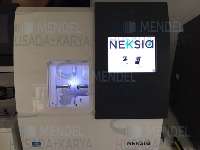

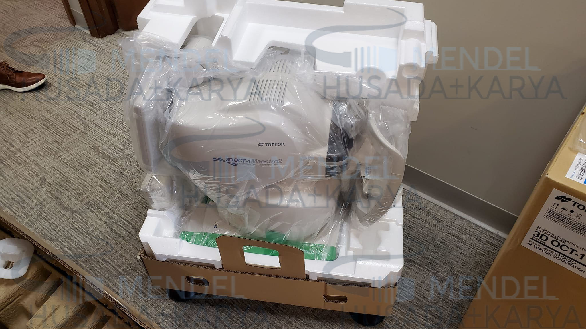
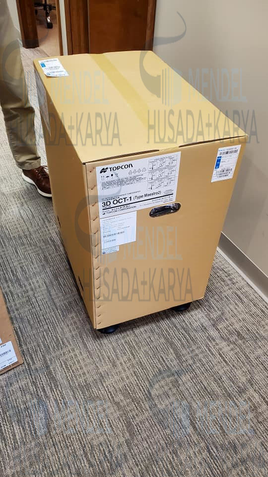
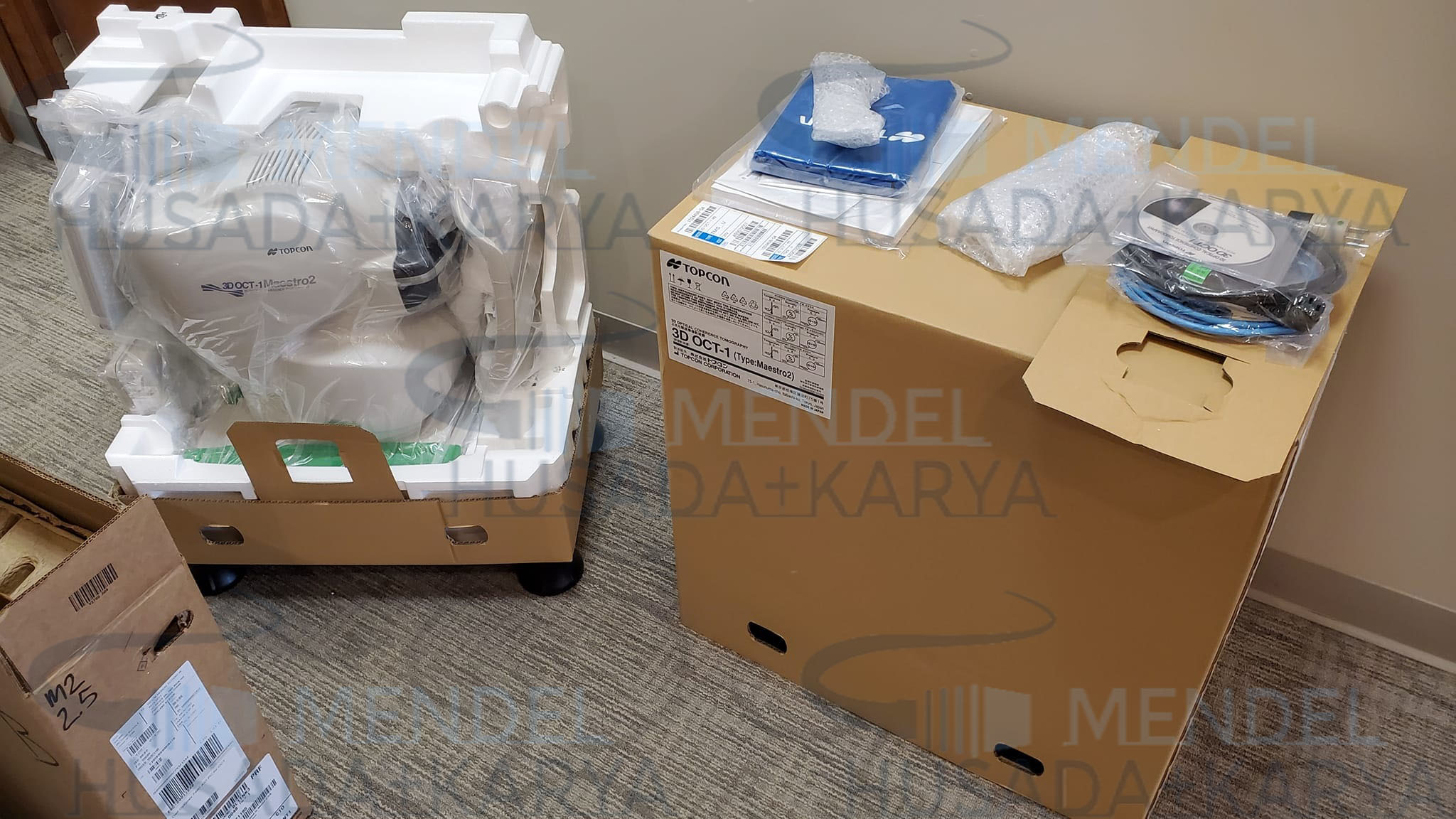
Topcon 3D OCT-1 Maestro 2
Original price was: $32,000.00.$15,490.00Current price is: $15,490.00.
Topcon 3D OCT-1 Maestro 2 is the user-friendly OCT-Fundus Camera system that automatically performs alignment, focus and capture with a single touch. The resulting reports enable comprehensive analysis of the macula, optic disc and anterior segment. Reports can be auto exported, quickly printed or sent to your EMR in common file formats.
Description
Topcon 3D OCT-1 Maestro 2
With the Maestro2, you have fast, multi-modality OCT/Fundus imaging, and advanced data management. The Maestro2 is the complete clinical workstation for any busy practice. With a single touch, the Maestro2 automatically performs alignment, focus, optimizing and capturing. After capturing, the report can be immediately displayed by clicking on the icon. In addition to automated capture, the Maestro2 offers manual/semi-manual options for difficult-to-image patients.
The Maestro2 has an integrated full-color fundus camera. With one touch, you can simultaneously acquire a posterior OCT image and a true color fundus image. This allows for PinPoint Registration and structural confirmation of the pathology. A small pupil function is also available, as well as fundus only capture.
Topcon 3D OCT-1 Maestro 2 Features :
– Combined OCT and color retinography
– Fully automated capture
– Compact, space-saving design
– Wide 3D scan and glaucoma module with Hood Report
– Normative database for retinal thickness, RNFL, ganglion cells and ganglion complex
– Automatic layer segmentation
– Anterior segment OCT
– Peripheral fundus photography
– 3D visualization
Topcon Healthcare empowers providers with advanced imaging, diagnostic solutions and intelligent data technology, offering a more fully integrated approach to diagnosis and treatment in eyecare.
| OBSERVATION & PHOTOGRAPHY OF FUNDUS: | |
| Type of photography | Color, Red-free* & IR*** |
| Picture angle of photography | 45° ± 5% or less |
| 30° or equivalent (digital zoom) | |
| Operating distance | 34.8 ± 0.1mm (when taking a picture of fundus) |
| Photographable diameter of pupil | Normal pupil diameter: ø4.0mm or more |
| Small pupil diameter: ø3.3mm or more | |
| Fundus image resolution (on fundus) | Center: 60 lines/mm or more |
| Middle (r/2) : 40 lines/mm or more | |
| Middle (r) : 25 lines/mm or more | |
| IR photography : Center: 5 lines/mm or more*** | |
| OBSERVATION & PHOTOGRAPHY OF FUNDUS TOMOGRAM: | |
| Scan range (on fundus) Horizontal direction | 3 – 12mm ± 5% or less |
| Vertical direction | 3 – 9mm ± 5% or less |
| Scan pattern | 3D scan (horizontal/vertical) |
| Linear scan (Line-scan/Cross-scan/Radial-scan) | |
| Scan speed | 50,000 A-Scans per second |
| Lateral resolution | 20µm or less |
| In-depth resolution | 6µm or less |
| Photographable diameter of pupil | ø2.5mm or more |
| OBSERVATION & PHOTOGRAPHY OF FUNDUS IMAGE / FUNDUS TOMOGRAM: | |
| Fixation target | Internal fixation target: |
| Dot-matrix type organic EL | |
| The display position can be changed and adjusted. | |
| The displaying method can be changed. | |
| Peripheral fixation target: | |
| This is displayed according to the internal fixation target displayed position. | |
| External fixation target | |
| OBSERVATION & PHOTOGRAPHY OF ANTERIOR SEGMENT: | |
| Type of photography | Color & IR |
| Operating distance | 62.6 ± 0.1mm (when taking a picture of anterior segment)** |
| OBSERVATION & PHOTOGRAPHY OF ANTERIOR SEGMENT TOMOGRAM: | |
| Operating distance | 62.6 ± 0.1mm (when taking a picture of anterior segment)** |
| Scan range (on cornea)** | Horizontal direction: 3 – 6mm ± 5% or less |
| Vertical direction: 3 – 6mm ± 5% or less | |
| Scan pattern | Linear scan (Line-scan/Radial-scan) |
| Scan speed | 50,000 A-Scans per second |
| Fixation target | External fixation target |
Related products
-
- Sale!
- Ophthalmology
ICare IC200 Handheld Tonometer
- Original price was: $1,590.00.$1,180.00Current price is: $1,180.00.
- Add to cart
-
- Sale!
- Medical Equipment, Ophthalmology
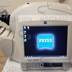
Zeiss Cirrus Hd-Oct 4000
- Original price was: $7,700.00.$4,280.00Current price is: $4,280.00.
- Add to cart
-
- Sale!
- Ophthalmology
Essilor Mr Orange Edger Machine
- Original price was: $14,290.00.$7,130.00Current price is: $7,130.00.
- Add to cart


Reviews
There are no reviews yet.