


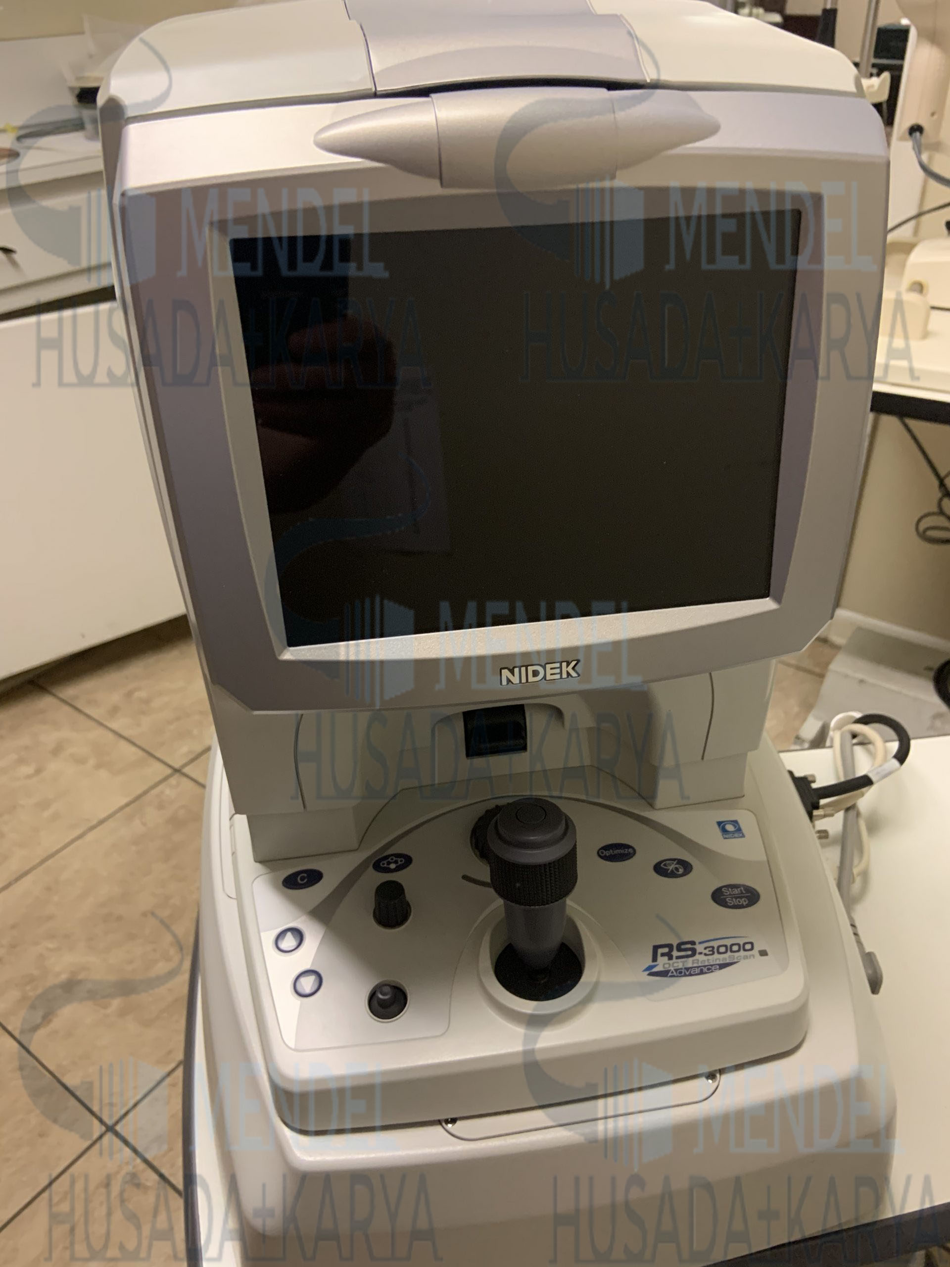
Nidek RS-3000 Advance 2 OCT
Original price was: $7,990.00.$3,720.00Current price is: $3,720.00.
The RS-3000 Advance 2 retains some of the signature features of the previous model including glaucoma analysis with a 9 x 9 mm wide area normative database, accurate image capture with a SLO based eye tracer, and selectable OCT sensitivity that allows acquisition of B-scan images through media opacities. Enhancements of the RS-3000 Advance 2 include 85,000 A-scans/s, 12 x 12 mm auto panorama imaging of OCT-Angiography, and increased image quality.
Description
Nidek RS-3000 Advance 2 OCT
Tracing HD Plus
The tracing HD plus function traces involuntary eye movements to maintain the same scan location on the SLO image for accurate image capture. This function allows accurate averaging of up to 120 images.
Nidek RS-3000 Advance 2 OCT FEATURES
OCT Angiography
53,000 Measurements/second
Torsion Eye Tracing
Scanning Laser Ophthalmoscope for Exceptional Retinal Imaging
HD Rastering of up to 120 Scans
6 Layer Segmentation
Wide Area Scan (12mm x 9mm)
Intuitive Glaucoma Analysis (RNFL)
Ganglion Cell (RNFL + GCL + IPL) Analysis
Retinal Thickness Analysis
Optional Anterior Segment Module
Tracing HD Plus
The tracing HD plus function traces involuntary eye movements to maintain the same scan location on the SLO image for accurate image capture. This function allows accurate averaging of up to 120 images.
| Real time compensation for eye movement with SLO-based eye tracer results in more accurate scans, ensuring higher image quality and maximum reproducibility |
Technical Details
Dimensions: 380 (W) x 524 (D) x 515 (H) mm
Weight: 34 kg
Power Supply: 100, 120, 230 V AC; 50/60 Hz
Power Consumption: 300 VA
Features
HD Tracing Plus function for precise tracing of scan location
Enhanced Image function for improved resolution of vitreous retinal images
Various scanning patterns including Macula Multi and Macula Radial
Macula Comparison capabilities to analyze chronological changes in retinal thickness
OCT en face imaging for comprehensive assessment of retinal layers
AngioScan function using OCT-Angiography for detailed visualization of retinal microvasculature
Select and Rescan Mode (SR Mode) for efficient image acquisition and lesion location selection
Vessel Density Map and Perfusion Density Map for quantitative analysis of retinal vessels
Foveal Avascular Zone (FAZ) autodetection and Shape Analysis for rapid assessment of retinal morphology
Tracking function for longitudinal assessment of vascular changes over time
Request for Quote
Related products
-
- Sale!
- Ophthalmology
ICare IC200 Handheld Tonometer
- Original price was: $1,590.00.$1,180.00Current price is: $1,180.00.
- Add to cart
-
- Sale!
- Ophthalmology
Topcon 3D OCT-1 Maestro 2
- Original price was: $32,000.00.$15,490.00Current price is: $15,490.00.
- Add to cart
-
- Sale!
- Ophthalmology
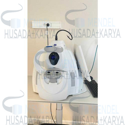
Retina Carl Zeiss Cirrus HD OCT 4000
- Original price was: $6,600.00.$4,210.00Current price is: $4,210.00.
- Add to cart


Reviews
There are no reviews yet.